
SP9 Protocol: Controlled Extraction Space Closure in Clear Aligner Therapy & Case Study
Written by Bushra Maayah, BDS

Introduction
Extraction space closure is one of the most biomechanically challenging aspects of clear aligner therapy. Aligners are highly effective for alignment, leveling, and mild to moderate movements. However, controlling mesiodistal bodily translation to close extraction spaces presents unique challenges.
These challenges include:
- Uncontrolled crown tipping
- Anchorage loss
- Reduced root control
To improve movement predictability and tipping control during extraction space closure, structured staging systems such as the SP9 (Shift–Pause–9) protocol are used. This blog explains what the SP9 protocol is, why it is necessary, and how it improves biomechanical control in aligner therapy.
What Is the SP9 Protocol?
SP9 stands for Shift–Pause–9. It is a staged movement protocol designed primarily for extraction space closure cases to control tipping in clear aligner therapy. The concept is simple and can be explained from the name itself:
- Shift: Teeth adjacent to the extraction space are actively moved or shifted for 9 steps towards the space
- Pause: Then, the same teeth remain stationary or pause for 9 steps while other teeth in the arch move
- The cycle repeats until space closure is achieved
→ The goal is to control tipping during mesiodistal translation. By pausing force application for 9 steps, crown displacement is ultimately reduced, allowing better root control and minimizing uncontrolled tipping. [6]
Why Is SP9 Needed in Extraction Cases?
Clear aligners primarily deliver force at the crown level. When attempting bodily movement into an extraction space, this can usually create a tendency for crown tipping toward the space, rather than controlled root translation.
Systematic reviews have shown that:
- Aligners are less predictable for bodily translation compared to tipping movements [1, 2]
- Complex movements require careful staging and attachment design to improve movement predictability [3]
In extraction cases, continuous mesiodistal movement without staging increases:
- Tipping moments
- Anchorage strain
- Risk of tracking loss
SP9 was developed to reduce these risks through controlled sequencing of movements.
How Does SP9 Improve Biomechanical Control?
- Tipping Control Through Pause Phases
During the shift phase, slight tipping may occur as the teeth adjacent to the extraction space translate or move to close the space. During the pause phase, the teeth adjacent to the extraction space remain stationary to allow:
- Gradual re-engagement of attachments
- Recovery of moment control
- Uprighting through aligner seating
- Anchorage Management
Moving the entire arch simultaneously increases anchorage demand and may reduce control. Segmented movement: [4]
- Distributes force demands
- Reduces plastic overload
- Improves tracking
Studies confirm that extraction space closure with aligners produces significant tipping forces if not properly staged. [1]
How Do Attachments Influence SP9 Success?
Attachment design plays a crucial role in moment control. Research shows:
- Attachment geometry affects force vectors and moment-to-force ratios. [1]
- Proper placement improves bodily movement predictability. [1]
- Poor staging combined with inadequate attachment design increases tipping risk. [2, 3]
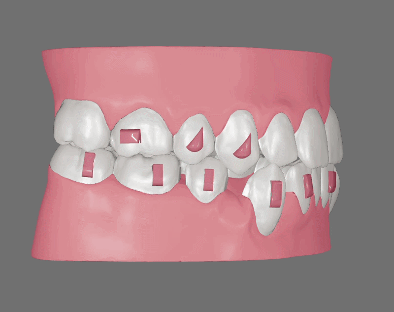
In extraction space closure, it is recommended to place vertical beveled rectangular attachments on teeth being translated to enhance root control and resist unwanted tipping. [5]
Case Study: SP9 Protocol Applied in a Class III Extraction Case
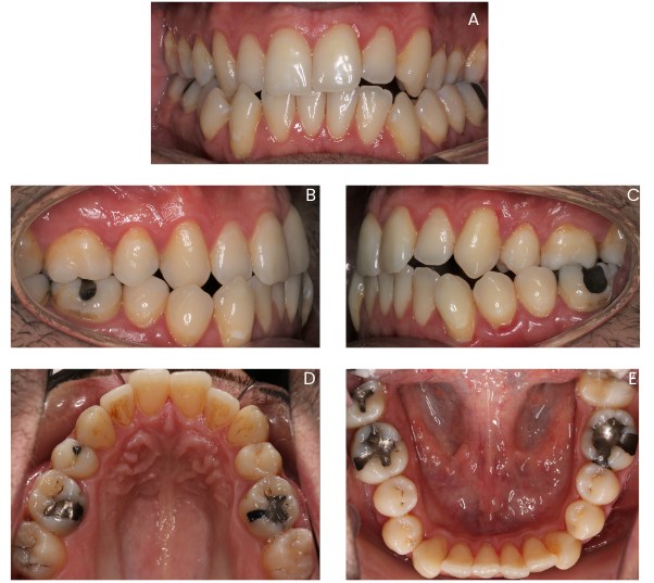
This is a case treated by the talented Dr. Salim Qadri using Eon Aligner. The patient was an adult male patient who presented with a Class III malocclusion requiring an extraction of the lower right first premolar followed by closure of the space. Dr. Qadri has requested to close that space using the SP9 protocol. As shown in figure 1 below, the before photos include the lower right first premolar, but it had been digitally extracted on the setup as requested by the doctor. The primary biomechanical concern for this case was the uncontrolled crown tipping during mesiodistal space closure.
One of the solutions was to include vertical beveled rectangular attachments to help with that concern. While attachments improve force application, they do not fully eliminate the risk of tipping when continuous translation is planned without proper staging and sequencing of movements. Therefore, to improve control and movement predictability, the SP9 protocol was incorporated into the treatment design.

As shown in the first 9 steps in figure 2 above, the teeth adjacent to the extraction space — the lower right canine and lower right second premolar — were translated toward the space in a controlled bodily manner. Vertical beveled rectangular attachments were placed on both teeth to enhance engagement and support root control during this phase.
In the following 9 steps, as shown in figure 3, these previously activated teeth remained stationary while other segments of the arch were moved. During this pause phase, the attachments continued to assist with uprighting and maintaining root position while anchorage demands were redistributed elsewhere in the arch.
.gif)
This sequence was then repeated as shown in figure 4. The lower right canine and second premolar were reactivated for another 9-step shift, followed by another pause phase, and so on, until complete space closure was achieved.
Protocol-in-Action
%20(1).gif)
.gif)
Figures 5 A & B demonstrate the entire sequence continuously, illustrating how the alternating shift-and-pause cycles progressed step-by-step until complete space closure was achieved from both the right & occlusal views.
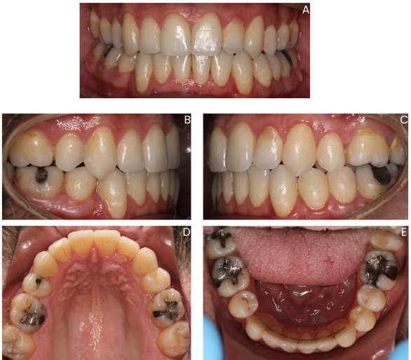
As shown in the post-treatment photos in Figure 6, the primary treatment objective was successfully achieved. The extraction space was fully closed without undesirable distal tipping of the lower right canine or mesial tipping of the lower right second premolar into the space. Maintaining this level of control also contributed to improvement of the anteroposterior relationship on the right side and supported a more stable interdigitation overall.
Although this technique may result in a longer treatment time, the result reflects the impact of thoughtful staging and proper sequencing of movements, rather than relying on continuous, uncontrolled translation. It also underscores the role of well-designed attachments in facilitating controlled space closure and maintaining root position throughout treatment.
Conclusion
Clear aligners inherently favor tipping over bodily translation during mesiodistal movement. A solution to this problem as proven by the case study shown is the adoption of the SP9 protocol when planning which introduces structured staging and sequencing to:
- Control crown angulation or tipping
- Improve attachment engagement
- Distribute anchorage demand
- Enhance movement predictability
It is not merely a sequencing method, it is a biomechanical tipping control strategy designed to improve results in extraction-based aligner therapy.
FAQS
References
Related Articles

Overcoming Common Challenges with Clear Aligners

Aligner Treatment Planning Software: What all Orthodontists Should Know

Bouncing Protocol: Controlled Deep Bite Correction in Clear Aligner Therapy
.svg)
© Eon Aligner Copyright 2023 All rights reserved